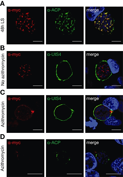
Fig. 4.

Apicoplast localization of PALM. A. Co-staining of fixed, PALM-mCherry-myc parasite-infected hepatoma cells 48 h after infection using anti-myc and anti-ACP antibodies. Note the substantial overlap between PALM and the signature apicoplast protein. B. PALM-mCherry-myc parasite-infected hepatoma cells left untreated, fixed at 48 h after infection, and stained with antibodies specific for the myc epitope and upregulated in infectious sporozoites protein 4 (UIS4), a signature protein of the parasitophorous vacuole. Note the branched PALM-positive structures. C. Antibiotics treatment with 1 µM azithromycin abolishes the branched PALM-positive structures while parasites appear to remain healthy otherwise. D. Antibiotics treatment with 1 µM azithromycin abolishes the branched PALM-positive structures as well as ACP-positive structures. All bars, 10 µm.
