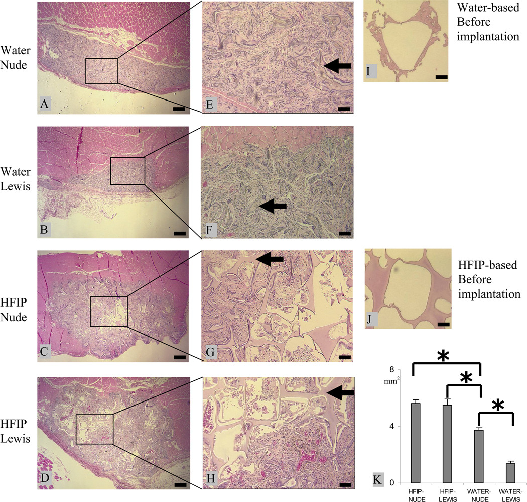
Figure 1.
Intramuscular degradation of aqueous-(A, B, E, and F) and HFIP-(C, D, G, and H) derived silk scaffolds in nude and Lewis rats. Scaffolds were implanted for 8 weeks and stained with H&E. Original structure of the aqueous- and HFIP-derived scaffolds prior to implantation are shown in I and J, respectively. The cross-section area is shown in K. Bars in A–D = 400 µm and in E–J = 100 µm. Solid arrows = remaining scaffolds. (p<0.05)

