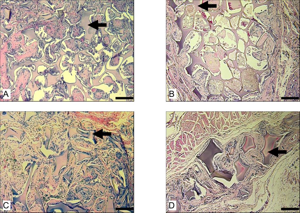
Figure 7.
Morphologies of subcutaneously implanted HFIP-derived silk fibroin scaffolds at 6 and 12 months. Images A and B are for HFIP-derived scaffolds prepared from 17% silk fibroin solution with 100 – 200 µm pore size at 6 months and 12 months, respectively. Images C and D are for HFIP-derived scaffolds prepared from 6% silk fibroin solution with 500 – 600 µm pore size at 6 and 12 months, respectively. Bars = 100 µm. Solid arrows = remaining scaffolds.

