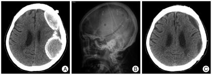
Fig. 3.
A : Preoperative computed tomography (CT) showing double CSDH with thickened calcified membrane. B : Postoperative lateral skull X-ray film showing large craniotomy on the left hematoma site. C : Postoperative CT showing total removal of hematoma with small residual subdural fluid collection. CSDH : chronic subdural hematoma.

