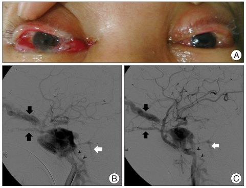
Fig. 1.
Preprocedural findings of a patient with bilateral carotid-cavernous fistulae. Photograph shows bilateral proptosis, chemosis, and eyelid edema (A). Right (B) and left (C) carotid angiograms reveal bilateral carotid-cavernous fistulae with shunts to the ophthalmic vein (black arrows), inferior petrosal sinus (black arrowheads), and petrosal vein (white arrow).

