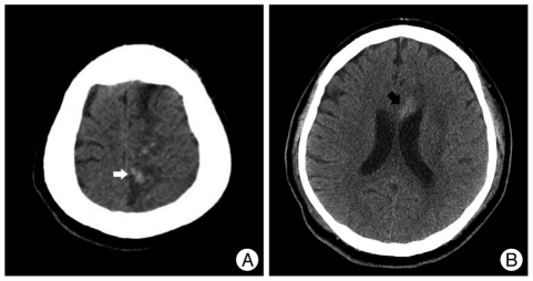
Fig. 3.
Follow-up CT images. One-day (A) and two-day (B) follow-up CT scans show two focal hemorrhagic foci in the left parietomedial region (white arrow) and the genu of the corpus callosum (black arrow), both of which occurred in the previously infarcted territory of the left anterior cerebral artery.

