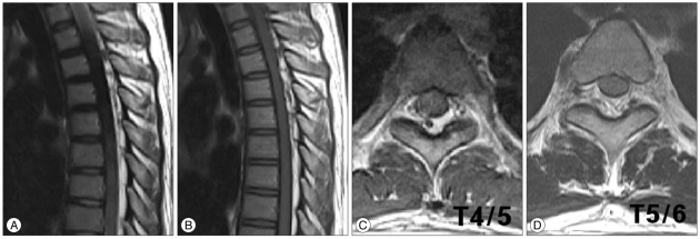
Fig. 3.
Thoracic spinal magnetic resonance imaging; Sagittal T2-WI (A), sagittal T1-WI (B), T4/5 axial T2-WI (C) and T5/6 axial T2-WI (D), showing a posterior epidural mass with mild compression of the dura extending from T3 to T9 segments with high signal intensity at T2-WI (A) and T1-WI (B) similar to that of subcutaneous adipose tissues. This epidural mass is extending into the left vertebral foramen on thoracic 4/5 (C), and T5/6 (D) levels. WI : weighted image.

