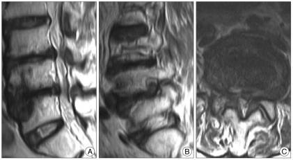
Fig. 1.
Preoperative magnetic resonance T2-weighted images. A : Midsagittal image showing the sclerotic change in both L4 lower and L5 upper endplates. B : Parasagittal image revealing the left L4-5 extraforaminal herniated disc. C : Axial image demonstrating the central stenosis on the level of the L4-5 disc space.

