Figure 1.
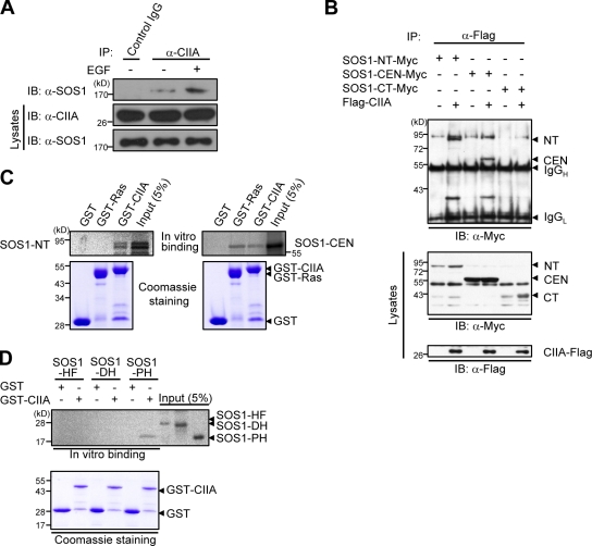
CIIA physically interacts with SOS1. (A) HeLa cells were deprived of serum for 16 h, incubated without or with 100 ng/ml EGF for 5 min, lysed, and subjected to immunoprecipitation (IP) with the anti-CIIA antibody or with control preimmune IgG. The resulting precipitates as well as the lysates were subjected to immunoblotting (IB) with antibodies to SOS1 or CIIA. (B) 293T cells were transfected for 48 h with various combinations of expression vectors for the indicated proteins. Cell lysates were immunoprecipitated with the anti-Flag antibody, and the resulting precipitates were examined by immunoblot analysis with the anti-Myc antibody. Cell lysates were also immunoblotted with antibodies to Flag or to Myc. IgGH and IgGL indicate heavy and light chains, respectively, of IgG. (C and D) In vitro–translated 35S-labeled SOS1 variants were pulled down with GST or GST-fused proteins immobilized on glutathione–agarose beads. The bead-bound 35S-labeled proteins were analyzed by SDS-PAGE and autoradiography. The gels were also stained with Coomassie brilliant blue. A fraction (5%) of the 35S-labeled protein input to the binding reaction is also shown. NT, N-terminal domain; CEN, central domain; CT, C-terminal domain.

