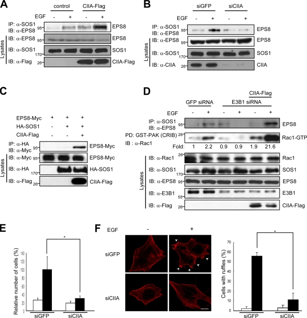
Figure 4.
CIIA promotes SOS1-mediated activation of Rac1. (A and B) MDCK/CIIA-Flag and MDCK/control cells (A) or HeLa cells expressing GFP (control) or CIIA siRNA (B) were deprived of serum for 16 h, incubated for 5 min with or without 100 ng/ml EGF, and lysed. Cell lysates were immunoprecipitated (IP) with the anti-SOS1 antibody, and the resulting precipitates were immunoblotted (IB) with the anti-EPS8 antibody. (C) 293T cells were transfected for 48 h with vectors for EPS8-Myc, HA-SOS1, and CIIA-Flag as indicated. Cell lysates were immunoprecipitated with the anti-HA antibody, and the precipitates were immunoblotted with the anti-Myc antibody. (D) HeLa cells were transfected with either GFP or E3B1 siRNA alone or together with a vector encoding CIIA-Flag. After 8 h of transfection, the cells were deprived of serum for 16 h and then incubated for 5 min with or without 100 ng/ml EGF. Cell lysates were immunoprecipitated with the anti-SOS1 antibody, and the resulting precipitates were immunoblotted with the anti-EPS8 antibody. Cell lysates were also subjected to pull-down (PD) with GST-CRIB and assayed for Rac1 activity. (E) HeLa cells stably expressing GFP or CIIA siRNA were deprived of serum for 16 h, transferred to the upper chambers of a Transwell plate, incubated for 24 h with (black bars) or without (white bars) 100 ng/ml EGF in the lower chambers, and analyzed for migration. (F) HeLa cells stably expressing GFP or CIIA siRNA were serum starved for 16 h, incubated for 5 min with (black bars) or without (white bars) 100 ng/ml EGF, and stained with Alexa Fluor red–conjugated phalloidin to detect F-actin. Arrowheads indicate F-actin–enriched membrane ruffles. The number of cells with ruffles were counted and expressed as the percentages relative to the total number of cells. Data are means ± SD from three independent experiments. *, P < 0.01. Bar, 10 µm.

