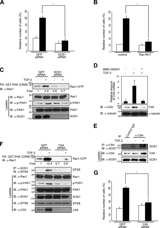
Figure 5.
CIIA mediates TGF-β–induced migration of A549 cells. (A, B, and G) A549 cells were transfected either with GFP (control), SOS1 (A), or CIIA (G) siRNA oligonucleotides or with a vector for Rac1N17 or the corresponding empty vector (B). After 8 h of transfection, the cells were deprived of serum for 16 h, transferred to the upper chambers of a Transwell plate, incubated for 48 h with (black bars) or without (white bars) 2 ng/ml TGF-β in the lower chambers, and analyzed for migration. Data are means ± SD from three independent experiments. *, P < 0.01. (C) A549 cells were transfected with GFP or SOS1 siRNA for 16 h, deprived of serum for 10 h, and incubated with or without 2 ng/ml TGF-β for 16 h. Cell lysates were then subjected to pull-down (PD) with GST-CRIB and assayed for Rac1 activity. (D) A549 cells were incubated with or without 10 µM BMS345541 for 1 h and then in the presence or absence of 2 ng/ml TGF-β for 6 h (for quantitative RT-PCR analysis of CIIA mRNA) or for 16 h (for immunoblot [IB] with antibodies to CIIA or α-tubulin [loading control]). RT-PCR data are means ± SD of triplicates from a representative experiment. (E) A549 cells were left untreated or treated with 2 ng/ml TGF-β for 16 h, lysed, and immunoprecipitated (IP) with the anti-CIIA antibody or rabbit preimmune IgG. The resulting precipitates were immunoblotted with the anti-SOS1 antibody. (F) A549 cells were transfected for 12 h with GFP or CIIA siRNA oligonucleotides, deprived of serum for 16 h, and incubated with or without 2 ng/ml TGF-β for 16 h. Cell lysates were then subjected to Rac1 activity assay as in C. Cell lysates were also immunoprecipitated with the anti-SOS1 antibody, and the resulting precipitates were immunoblotted with the anti-EPS8 antibody.

