
Figure 2.
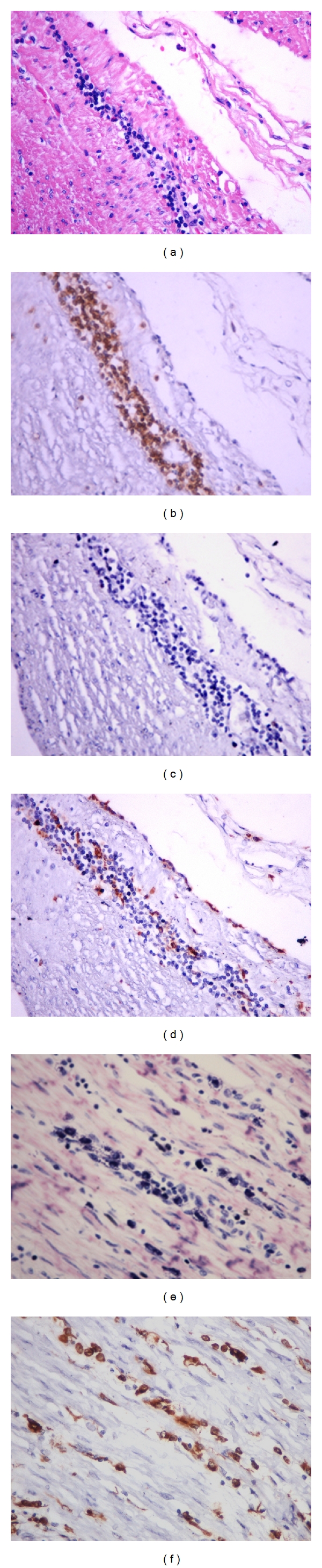
(a)–(d) Myositis in esophagus without mega. Note that the infiltrate is predominantly mononuclear ((a) HE—400x), with intense predominance of T lymphocytes ((b) immunohistochemistry for CD3—400x); extremely rare B lymphocytes ((c) immunohistochemistry for CD20—400x), and few positive CD68 cells ((d) immunohistochemistry for CD68—400x). (e) Numerous mast cells in the perimysium and in the foci of myositis in a case of megaesophagus can be seen (Giemsa—400x). (f) Compare this section stained by immunohistochemistry for CD68 with the previous figure, showing that both macrophages and mast cells are CD68 positive (400x).