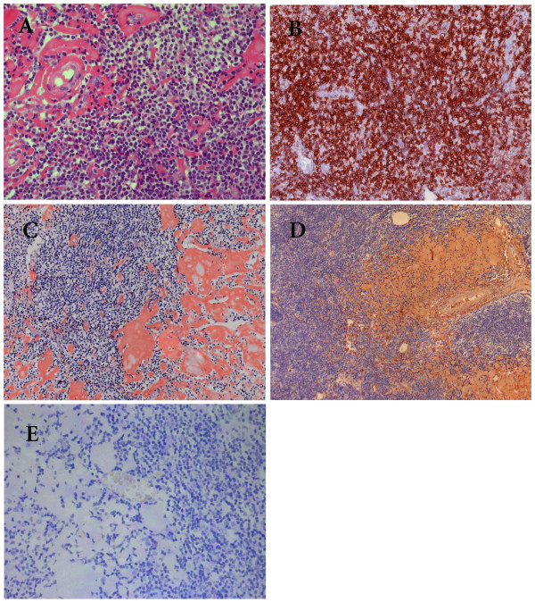
Figure 3.
Photomicrographs of histological changes of the affected lymph node. (A). Lymph node involved showing diffuse small lymphocyte-like cells and patches of homogenous materials in the dark background of proliferating cells. (HE staining×400). (B) The proliferating cells are CD5 positive by immunohistochemistry (×200). (C). The deposit was strongly Congon-Red positive. (×200). (D). Strong positivity of κ light chain demonstrated by immunohistochemistry (×100). (E). The deposit was completely negative for λlight chain. (×200).

