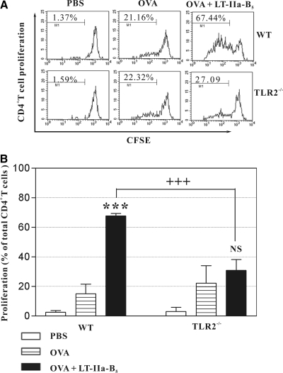
Figure 5. i.n. administration of LT-IIa-B5 induces enhanced Ag-specific proliferation of CD4+ T cells in the CLN.
Syngeneic CFSE-labeled DO11.10 CD4+ T cells (2×10⁶) were adoptively transferred into WT BALB/c or BALB/c(TLR2−/−) mice. Subsequently, mice were i.n.-immunized with OVA (control) or with a combination of OVA and LT-IIa-B5. Three mice were used for each group. CLNs were removed at Day 3 after immunization, and total cells were stained with allophycocyanin-anti-mouse DO11.10 and PerCP-anti-mouse CD4. CFSE fluorescence of the DO11.10+CD4+ T cells was measured as a determinant of proliferation. (A) Histograms of the proliferative responses of CFSE-labeled DO11.10 CD4+ T cells in the CLN. In each histogram, the percentage of dividing cells in each population is denoted above the bracket. Data are shown from one of three independent experiments, each of which yielded consistent results. (B) Graphical comparison of the percentage of dividing CFSE-labeled DO11.10 CD4+ T cells in the CLN. Error bars denote 1 sd from the mean for groups of three mice. Data from one of two independent experiments, each of which yielded consistent results, are shown. WT, TLR2-proficient BALB/c mice; TLR2−/−, TLR2-deficient mice; ***statistical difference from the control (OVA; P<0.001); +++statistical difference (OVA+LT-IIa-B5-treated, WT vs. TLR2-deficient; P<0.001); NS, OVA versus OVA + LT-IIa-B5, TLR2-deficient mice.

