
Figure 3.
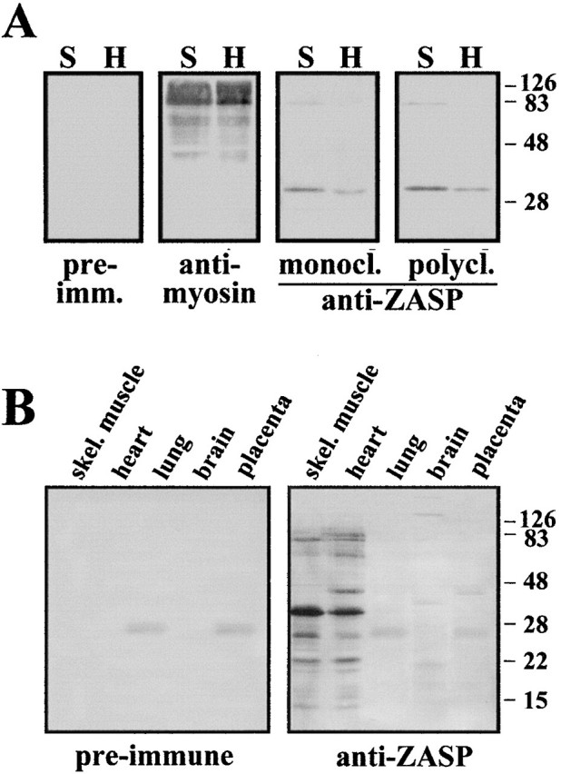
(A) Western blot analysis of heart and skeletal muscle tissue with antibodies to myosin, ZASP, and preimmune sera. Equal amounts of proteins were run in each lane (10 μg) on a 15% SDS-polyacrylamide gel and then blotted onto Immobilon-P membrane. The ZASP mAb was used undiluted, whereas the pAb was used at 1/20,00 dilution, as were the preimmune and myosin sera. (B) Tissue distribution of ZASP as demonstrated by Western blotting. Protein extracts from human heart and skeletal muscle (10 μg), as well as from brain, lung, and placenta (60 μg) were loaded in each lane, run on a 15% SDS-polyacrylamide gel, and then blotted. The membrane was probed with mouse pAb specific for ZASP and preimmune mouse sera, both used at 1/200 dilution. Sigma Chemical Co. color molecular weight markers were used.