
Figure 3.
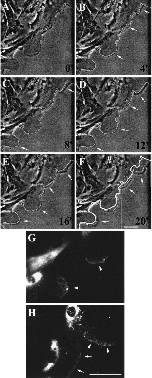
MMP-9 within migrating HBEC accumulates at the tip of extending lamellipodia. 1 d after injuring primary and confluent cultures of HBEC, a series of phase-contrast images of cells migrating and extending lamellipodia at the edge of the wound were taken at 4-min intervals (A–F). In images B–F, the leading front of the cells from the previous image (4 min earlier) has been highlighted with a thin white line. Comparison of this line to the present position of the cells enables the parts of the cells that have advanced during the 4-min period to be visualized (B–F, arrows). In F, the thick white line indicates the initial position of the cells shown in A. At the end of the experiment, the cells were fixed and MMP-9 distribution was assessed by immunofluorescence. (G and H) High magnification of MMP-9 distribution in the areas analyzed for cell movement (F, insets). MMP-9 is detected at the forefront of migrating cells with more intense fluorescence in more rapidly moving lamellipodia (G and H, arrowheads) than in those moving more slowly (H, arrows). Bars, 50 μm.