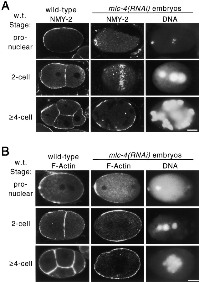
Figure 2.
Distribution of F-actin and nonmuscle myosin in wild-type and mlc-4(RNAi) embryos. Images were obtained by confocal microscopy (see Materials and Methods). (A) Distribution of NMY-2, a nonmuscle myosin II required for cytokinesis and polarity in the early embryo (Guo and Kemphues 1996). (first column) NMY-2 distribution in wild-type pronuclear, two-cell, and four-cell embryos. NMY-2 is uniformly localized to the cortex of early embryonic cells. (second column) NMY-2 distribution in mlc-4(RNAi) embryos. NMY-2 accumulates at the presumptive sites of cleavage furrow formation. At the pronuclear stage, a ring of NMY-2 localization is seen around the location of the polar body DNA (see third column for DNA localization; this localization was seen in 3/3 additional embryos scored at this stage). At the equivalent of the two-cell stage (middle), a surface focal plane image of the embryo shows a band of NMY-2 localization that extends around the circumference of the embryo (7/7 additional embryos at this stage showed similar localization). In later stages, shown in the bottom, NMY-2 is localized to bands that appear as patches in a confocal section. (third column) DNA localization in the same embryos as in column 2, as revealed by staining with DAPI. In the middle, note the extra, smaller, nuclear structure formed from the polar body chromosomes. (B) Distribution of F-actin revealed by rhodamine-conjugated phalloidin staining. (first column) F-actin distribution in wild-type pronuclear, two-cell, and four-cell embryos. F-actin appears uniformly distributed in the cortex of the embryonic cells. (second column) F-actin distribution in mlc-4(RNAi) embryos equivalent in age to pronuclear, two-cell, and four-cell embryos. As in wild type, F-actin is enriched at the cortex of the embryo. (third column) DNA localization in the same embryos as in column 2, as revealed by DAPI staining. Despite the localization of NMY-2 and F-actin to presumed regions of the cytokinetic furrow, no membrane ingression is seen in mlc-4(RNAi) embryos during mitotic cell cycles. Bars, 10 μm.

