Figure 3.
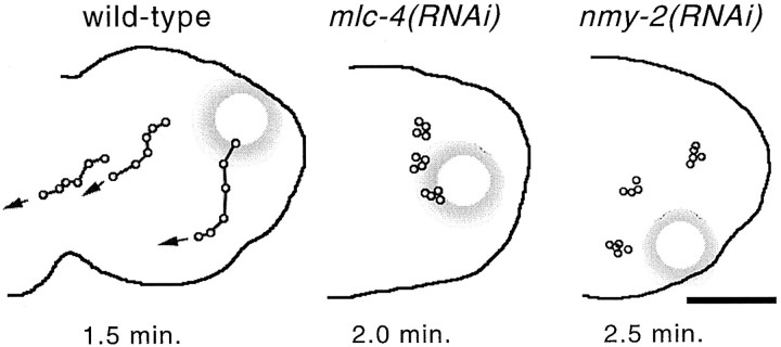
Cortical movement of yolk granules in wild-type, mlc-4 (RNAi), and nmy-2(RNAi) mutant embryos. Individual yolk droplets were followed by time-lapse digital photomicroscopy (see Materials and Methods) and were traced onto an outline of the respective embryos. Each dot along a line represents the position of the same yolk droplet at consecutive time intervals; the total elapsed time is shown below each embryo. The large shadowed circles represent the position of the paternal pronucleus in each embryo. (left) Cortical flow in a wild-type embryo. Note the cortical movement away from the paternal pronucleus. Analysis of 20 droplets in four different embryos yields an average speed of cortical yolk droplets at 4.5 ± 0.6 μm/min. (middle and right) Cortical yolk droplet movement in mlc-4(RNAi) and nmy-2(RNAi) mutant embryos, respectively. In these and three other embryos for each mutant, no directed yolk droplet movement was detected (20 granules for each mutant). Bar, 10 μm.

