Figure 7.
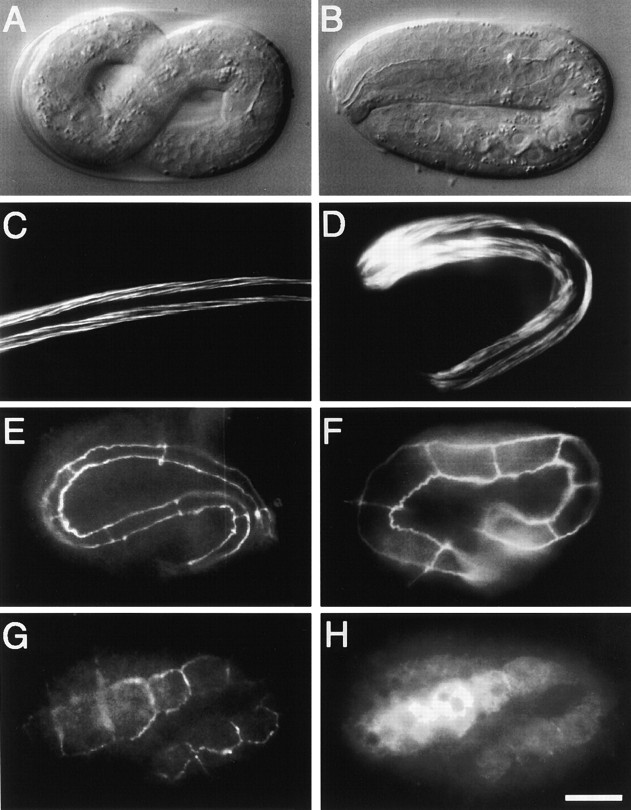
Embryonic elongation phenotype of mlc-4(or253). (A) Nomarski photomicrograph of a wild-type embryo just before hatching (12 h at 22°C). (B) Photomicrograph of an mlc-4(or253) embryo of similar age as in A. Note that the mutant embryo has only elongated to the twofold stage, whereas the wild-type embryo in A has elongated to the threefold stage (stages as in Wood 1988). mlc-4(or253) embryos do not elongate further and hatch as shortened, thicker larvae. (C) Fluorescence micrograph of phalloidin staining of actin in a wild-type larva shortly after hatching. Two of four bands of bodywall muscles are shown (micrograph is the same magnification as A, but the larva is no longer folded into the eggshell). (D) Phalloidin staining of actin in an mlc-4(or253) larva. Two of four bands of bodywall muscles are shown. Although the muscles appear structurally similar to wild type, the larva is shorter and thicker than wild-type larva. (E) Fluorescence micrograph of MH27 antibody staining in a wild-type larva of similar age as A. The epitope recognized by MH27 marks the boundaries of hypodermal cells. The focal plane shows 7 of 10 seam cells present along one side of the larvae. (F) MH27 staining in an mlc-4(or253) embryo of similar age to A, B, and E. 8 of 10 seam cells along one side of the larva are shown. Note that the seam cell morphology in the mlc-4(or253) embryo lacks the thin, elongated, appearance of seam cells in the wild-type embryo. (G) MH27 staining in a bean stage embryo that harbors an mlc-4::GFP expression construct and is just beginning the elongation process. Shown are 8 of 10 seam cells. (H) Same embryo as in G, double stained with antibodies recognizing GFP. Note that the anti-GFP antibody stains the seam cells, as revealed by comparison to the MH27 staining in G. All photomicrographs are at equivalent magnification. Bar, 10 μm.

