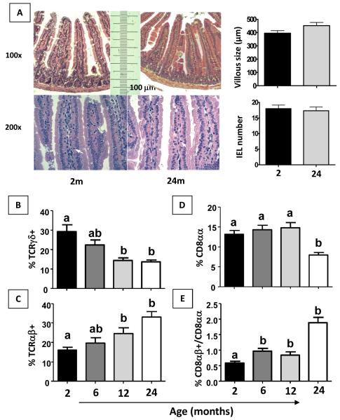
Figure 1. TCRγδ +, TCRαβ +, CD8αβ+ and ratio CD8αβ/CD8αα IELs are altered in aged mice.
Histology of H&E stained-sections from small intestine of 2 and 24-month-old non-manipulated mice. Villous length and IEL number were evaluated. Original magnification: 100x and 200x (A). Flow cytometry and frequency of IEL isolated from 2- to 24-month old non-manipulated mice stained with fluorescent antibodies to TCRαβ+, TCRγδ+, TCRαβ+CD8αα+ and TCRαβ+CD8αβ+ were gated in total lymphocytes (B-E). Bars represent the mean ± SEM of 5 mice per group. Letters (a, b) represent differences among groups (p< 0.05).

