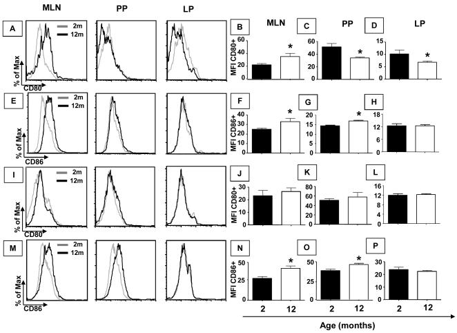
Figure 4. Expression of co-stimulation molecules CD80/86 in F4/80 and CD11c+ cells is affected by aging.
Mesenteric lymph nodes, Peyer’s patches and small intestine lamina propria cells were obtained from non-manipulated mice from 2 and 12 months of age. Histogram of CD80 (A) and CD86 (E) gated in F4/80+ cells as well as CD80 (I) and CD86 (M) gated in CD11c+ cells. Bars show the median (MFI) of cells stained with fluorescent antibodies anti-CD80 (B, C and D) gated in F4/80+ cells, anti-CD86 (F, G and H) gated in F4/80+ cells, anti-CD80 (J, K and L) and anti-CD86 (N, O and P) gated in CD11c+ cells. Median (MFI) of double positive cells, gated in total granulocytes, was assessed by flow cytometry. Bars represent the mean ± SEM of 5 mice per group (*p< 0.05).

