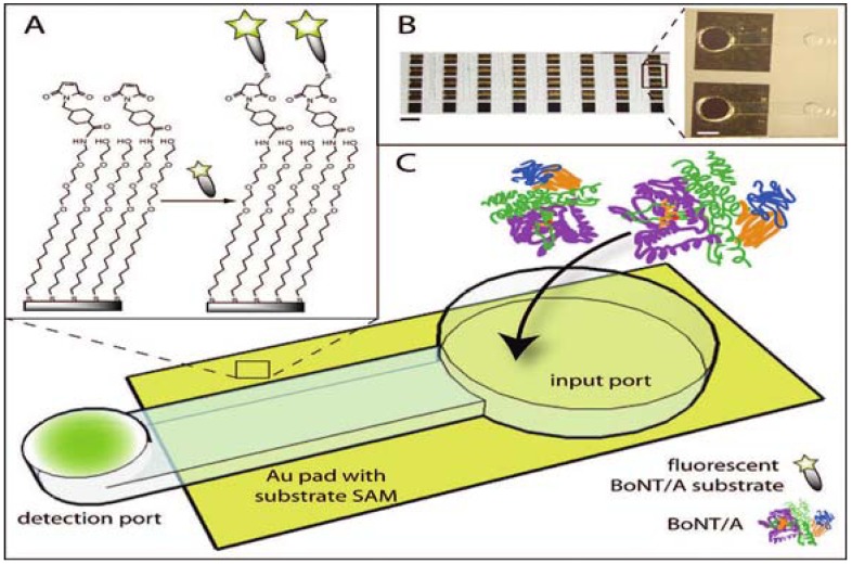
Figure 6.
Microfluidic device for fluorescent endopeptidase assay. (A) Fluorescein labeled BoNT/A substrate attached through the linker to the gold surface. (B) View of the array of 40 devices (scale bar = 5 mm). (C) BoNT/A is added into input port. During incubation, immobilized substrate is cleaved and the fluorescent fragment is released into the solution and concentrated at the detection port via evaporation. Reprinted with permission from [108]. Copyright © 2009 American Chemical Society.

