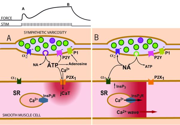
Figure 4. Scheme of Ca2+ signaling induced by sympathetic neuromuscular transmission in a small artery.

(A) Early during a train of nerve fiber action potentials, smooth muscle contraction is activated mainly by post-junctional Ca2+ transients (jCaTs) induced by neurally released ATP. JCaTs are localized to the post-junctional region, and arise from Ca2+ that has entered via P2X receptors. At this time, sympathetic varicosities may release mainly small vesicles that contain a relatively high concentration of ATP (viz. the relatively few ‘big’ quanta proposed by Stjarne, 2001) (B) Later during a train of nerve fibre action potentials, jCaTs are rare, and contraction is activated by Ca2+ waves that arise from sarcoplasmic reticulum (SR). Ca2+ release from SR is activated by InsP3, produced after binding of NA to α1-adrenoceptors. At this time, sympathetic varicosities may release small synaptic vesicles (the more numerous ‘small’ quanta, green) that contain a relatively high concentration of NA. An effect not shown in the scheme is the action of neurally released NPY to increase the frequency of NA induced Ca2+ waves. Figure reproduced from (Wier, 2003)).
