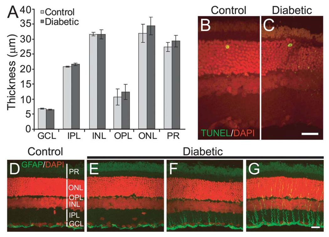
Fig. 2. Few overt signs of retinopathy are seen in the diabetic retina.
A: Mean thickness of retinal layers is not reduced in diabetic animals. GCL, ganglion cell layer; IPL, inner plexiform layer; INL, inner nuclear layer; OPL, outer plexiform layer; ONL, outer nuclear layer; PR, photoreceptors. B,C: Cell death was not increased in diabetic retinas. Very few TUNEL-positive cells (green/yellow profiles) were observed in both control (B) and diabetic (C) retinas. DAPI-labeled cell nuclei are shown in red. D–G: Immunostaining shows the expression of GFAP (green) in control (D) and three diabetic (E–G) retinas. A range of GFAP expression was observed in the vertically oriented Müller cells in diabetic retinas. Retinas from some animals were similar to controls (E), some showed a minor increase (F) while some showed substantial upregulation (G). GFAP-positive astrocytes, located beneath the GCL, are seen in both control and diabetic retinas. DAPI-labeled cell nuclei are shown in red. Scale bars, 20 µm.

