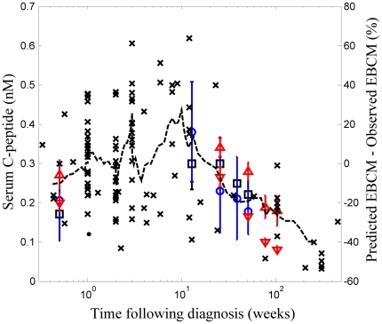
Figure 3. Dynamic change in residual beta cell mass corresponds to the dynamic change in plasma C-peptide following onset of type 1 diabetes.
The difference between predicted and observed excess beta cell mass (right axis: x) and plasma C-peptide (left axis: square [17], circle [18], ▵- children initially negative for autoantibodies at diagnosis and during follow-up [16], and ▿- children positive for at least one autoantibody [16]) shown as a function of time following clinical diagnosis of type 1 diabetes. Plasma C-peptide levels are reported as a mean + SE. A 9-point moving average of the difference in excess beta cell mass is shown for comparison (dotted line). The dynamic change in observed beta cell mass was obtained from pancreata obtained from patients with type 1 diabetes [9]–[11]. The predicted beta cell mass is an estimate of the minimum beta cell mass required to maintain glucose homeostasis.

