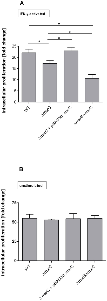
Figure 7. Intracellular proliferation of S. Typhimurium wild type, ΔmsrC, ΔmsrB .
ΔmsrC in RAW 264.7 cells. S. Typhimurium wild type, ΔmsrC, ΔmsrC + pBAD30::msrC and ΔmsrBΔmsrC were tested A. in IFN-γ-activated, and B. in non-stimulated RAW 264.7 cells. Intracellular proliferation was determined in a gentamicin protection assay. Number of bacteria 16 h post infection was divided by the number of bacteria 2 h post infection. Results shown are the means ± standard error of the mean for three independent experiments, each in triplicate. P-values <0.05 were considered to be significant as indicated by asterisks.

