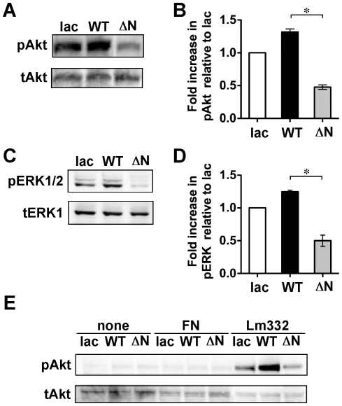
Figure 4. Effects of ΔNß4 integrin on cellular signaling.
Lac, WT or ΔN keratinocytes were grown to semi-confluence and then each cell lysate was collected as described in “MATERIALS AND METHODS. ” (A and C) 20 µg of cell lysates from those keratinocytes were run on 7.5% or 5–20% SDS-polyacrylamide gel and probed with a phospho-Akt Ab (A, upper panel) or phospho-ERK1/2 Ab (C, upper panel), and then reprobed with a total Akt Ab (A, lower panel) or total ERK Ab (C, lower panel), respectively. (B and D) Results of the densitometric analysis are shown as the integrated density of the ratio of phospho-protein to total protein bands, which was 1.0 for lac keratinocytes. *p<0.001 (one-way ANOVA, Bonferroni post test for WT) vs. ΔN. (E) Cells in supplement-free media were plated to the indicated substrates. After 30 minutes incubation, cell lysates were collected and immunoblotted against the indicated antibodies. All blots are representative of at least three independent experiments: none, plastic; FN, fibronectin; and, Lm332, laminin-332.

