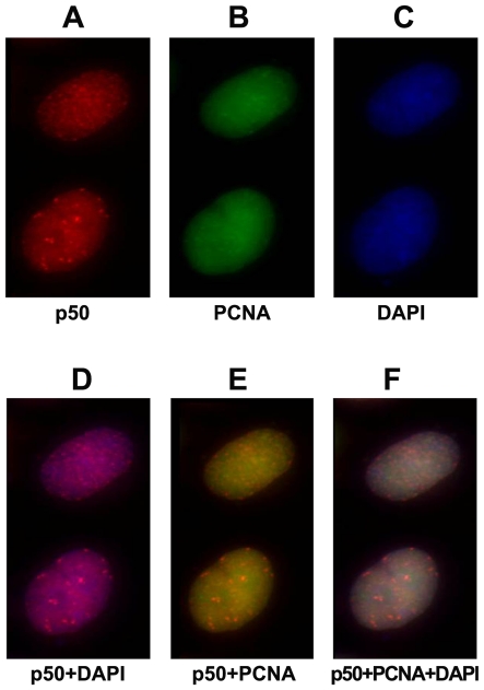
Figure 7. Co-nuclear staining of p50 and PCNA.
Hela cells were fixed, permeabilized, and co-stained for p50 and PCNA using indirect immunofluorescence in which an anti-p50 rabbit polyclonal antibody was used for p50 while an anti-PCNA mouse monoclonal antibody PC-10 was used for PCNA. A: Rhodamine-X-conjugated anti-rabbit IgG secondary antibody was used for p50 (red). B: Alexafluor488-conjugated anti-mouse IgG secondary antibody was used for PCNA (green). C: DNA was counterstained with DAPI immunofluorescence (blue). D: Merger for p50 (panel A) and DAPI (panel C). E: Merger for p50 (panel A) and PCNA (panel B). F: Merger for p50 (panel A), PCNA (panel B), and DAPI (panel C). Cells were analyzed and photographed with an Axiovert 200 M fluorescence microscope (Zeiss).

