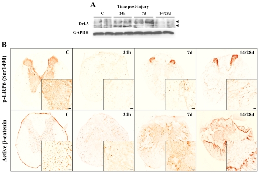
Figure 4. Active canonical Wnt signalling pathway in adult spinal cord before and after SCI.
(A) Western blot of Dvl-3 with protein samples isolated from a 1 cm long spinal cord fragment from non-lesioned control animals (C) and SCI animals at 1, 7 and 14/28 dpi. SCI induced hyperphosphorylation (upper arrowhead) and thus, the activation of Dvl-3 24 hours post-injury, which peaked at 7 dpi. GAPDH levels were used as a control for protein loading. (B) Representative histological images of the expression of the active LRP6 canonical co-receptor (phosphorylated at serine 1490) and β-catenin (dephosphorylated at serine 37 and threonine 41) before SCI and at 1, 7 and 14/28 dpi. Active LRP6 and β-catenin were both expressed in the grey matter of the non-injured spinal cord, the latter exhibiting a vascular-like pattern. After SCI, active LRP6 and β-catenin was expressed in cells located in the white matter of the wound epicentre, with a clear increase from 7 dpi and a final location suggestive of a role in tissue response. Scale bars = 50 µm.

