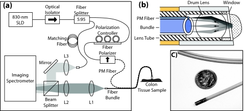
Figure 1.
a/LCI system diagram. (a) System illustration, (b) probe tip assembly illustration showing delivered (dark gray) and scattered (light gray) light, and (c) photograph of the endoscopic probe pictured next to a U.S. dime for scale. Illustration adapted from Zhu et al. (Ref. 22).

