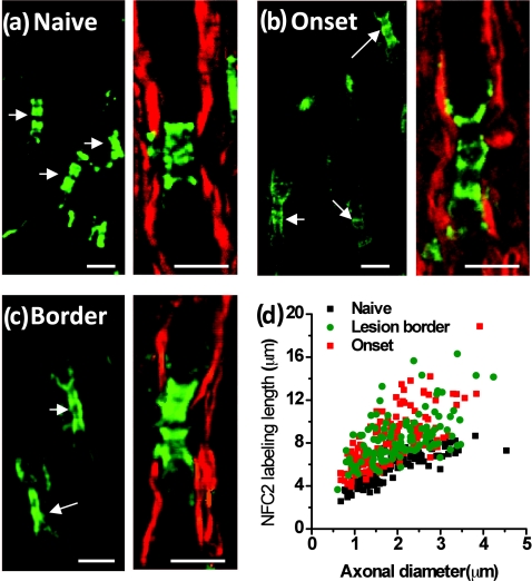
Figure 7.
Elongated neurofascin distribution in the paranodal domain. Green: immunolabeled NFC2 imaged by TPEF; red: myelin imaged by CARS. (a–c) Representative images of labeled NFC2 representing neurofascin 155 at the paranodal myelin and neurofascin 186 at the nodal axolemma in the white matter of naïve, EAE onset, and lesion borders at peak acute EAE. Elongated distribution of NFC2 was observed with extension into the internodes at locations no more than 100 μm away from the meninges at EAE onset and within lesion borders at peak acute EAE. (d) Comparison of NFC2 labeling lengths versus axonal diameters at different stages. Bar = 5 μm.

