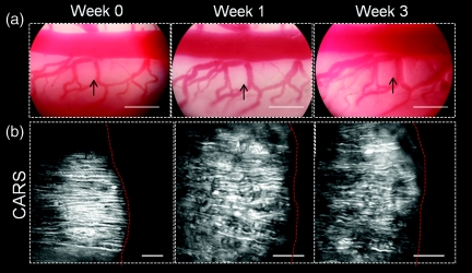
Figure 2.
Longitudinal photography and CARS imaging of the spinal cord white matter in a live rat. (a) Photographs taken at the eyepiece of an upright microscope recorded the general conformation of blood vessels as a landmark following 3 weeks. Arrowheads show the imaging area in (b). Bar = 500 μm. (b) CARS imaging did not show significant myelin damage over the 3 weeks in a rat injected with saline beneath dura. Right to the red dashed lines is the adjacent blood vessel. Bar = 20 μm.

