Figure 3.
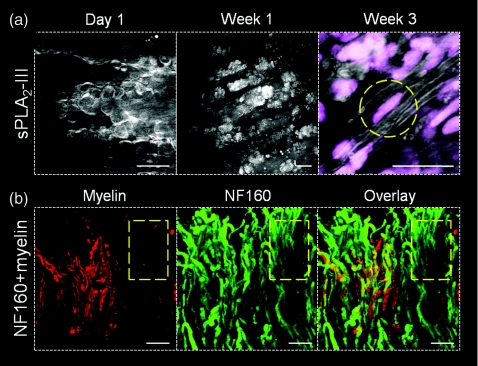
Longitudinal CARS imaging of sPLA2-III induced demyelination and spontaneous remyelination in a live rat spinal cord. (a) After sPLA2-III injection, myelin degradation was observed in 24 h by formation of myelin vesicles. These vesicles appear to be digested by macrophages/microglia 1 week after injection. By 3 weeks post-injection, signs of Schwann cell mediated remyelination were visible, with elongated cell nuclei (dashed circle) adjacent to myelinated axons at the injection site. (b) At 1 week post sPLA2-III injection, the myelin sheath was visualized by CARS (red), axons were visualized by immunofluorescent staining of NF160 (green). The overlay image showed the absence of myelin sheath (dashed square). Bar = 20 μm in all images.

