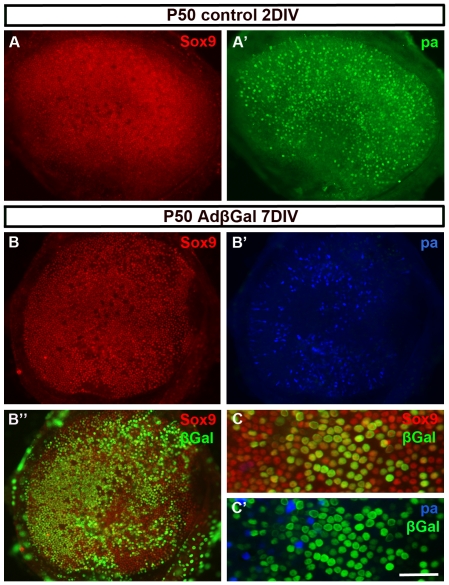
Figure 1. Adenoviruses infect adult utricular supporting cells in vitro.
(A, A') P50 explants maintained for 2 DIV show a homogenous layer of Sox9+ supporting cells (A) and parvalbumin+ hair cells (A'), based on double-immunofluorescence. (B-B″) At 7 DIV, triple-immunofluorescence reveals that the homogenous layer of Sox9+ supporting cells is maintained, but there is a loss of a large part of parvalbumin+ hair cells. β-galactosidase staining shows that supporting cell infectivity is more than 50% (B″). (C,C'). This higher magnification view from an explant triple-stained for Sox9, β-galactosidase and parvalbumin shows transgene expression in a large part of supporting cells, in contrast to remaining hair cells. Abbreviations: pa, parvalbumin; βGal, β-galactosidase. Scale bar, shown in C': A–B″, 140 µm; C,C', 60 µm.

