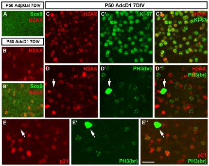
Figure 6. Induction of DNA damage in cell cycle reactivated supporting cells of the adult utricle. Absence of DNA damage and p21Cip1 expression in mitotic supporting cells.
(A) Sox9+ supporting cells in AdβGal-infected explants do not express the DNA damage marker p-H2AX. (B,B') In contrast, several Sox9+/p-H2AX+ supporting cells are present at 7 DIV in explants transduced by AdcD1. (C-C″) Double-immunofluorescence shows that p-H2AX expression is restricted to Ki-67+ cells. (D-D″) p-H2AX is expressed in late G2 cells, based on the staining pattern obtained with the “PH3-Ser10 broad antibody” (see text). Observe that cells with PH3+ mitotic profiles lack p-H2AX foci (arrows in D',D″). (E-E″) In AdcD1-infected explants at 7 DIV, p21Cip1 is expressed in late G2 cells, based on double-staining with the “PH3-Ser10 broad antibody”. Note that PH3+ mitotic profiles do not show p21Cip1 staining (arrows in E',E″). Abbreviations: βGal, β-galactosidase; cD1, cyclin D1; PH3(na), phosphorylated histone H3-Ser10 narrow; PH3(br), phosphorylated histone H3-Ser10 broad; H2AX, phosphorylated histone H2AX. Scale bar, shown in E″: A-B', 40 µm; C-E″, 30 µm.

