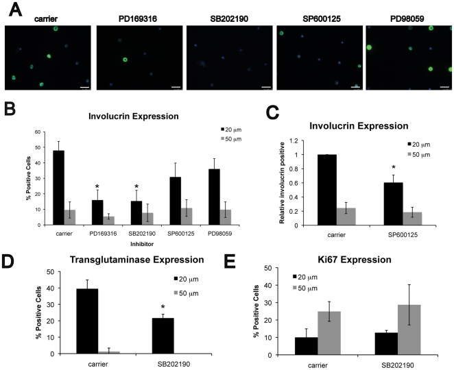
Figure 1. Effect of MAPK inhibitors on shape-induced terminal differentiation.
(A) Representative images of keratinocytes on micro-patterned substrates comprising 20 µm diameter collagen islands. Cells were cultured for 24 h in medium containing carrier (0.1% DMSO), 10 µM PD169316, 2 µM SB202190, 10 µM SP600125, or 10 µM PD98059. Immunofluorescence staining for involucrin (green) shows terminally differentiating cells, and nuclei are labeled with DAPI (blue). Scale bar = 50 µm. Quantification of (B-C) involucrin, (D) transglutaminase I, and (E) Ki67 positive cells after 24 h on 20 µm or 50 µm substrates. In (C), the proportion of involucrin positive cells was normalized to carrier-treated cells on 20 µm islands. Data are means ± SEM of 3 experiments. *P<0.05 compared to carrier.

