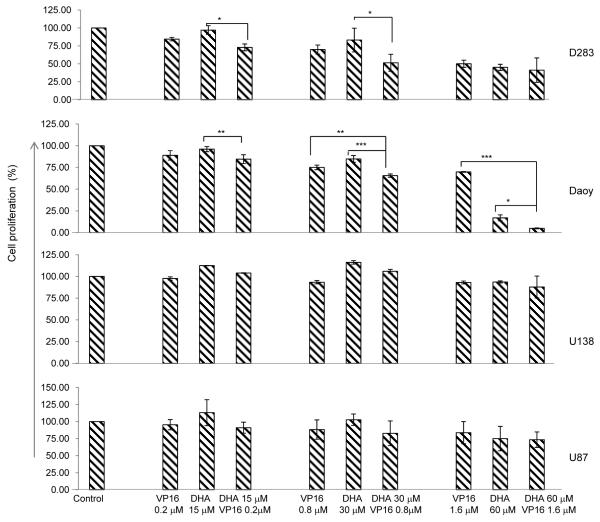
Fig. (1). The effects of DHA or VP16 alone and their combination on cell proliferation in brain tumor cells.
Cells in complete medium were placed in 96-well plates overnight before treatment. After 48 hours, the number of viable cells in each well was determined using MTS. The optical densities were measured at 490 nm. The results were calculated as the percentage of control cultures and presented as mean ± SD. The statistical differences were determined using paired student’s t-test and performed using SPSS. * p<0.05, **p<0.01, and ***p<0.001.

