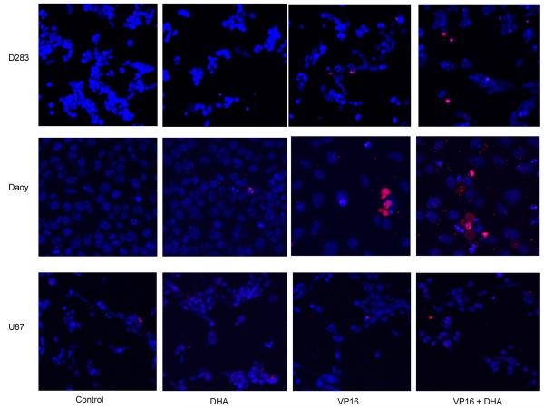
Fig. (2). The effects of cytotoxicity of DHA or VP16 alone and their combination in brain tumor cells.
The cells were seeded in 6-well plates for treatments. After 24 hours, the cells were stained in culture with Hoechest 33342/PI. Images were captured using confocal microscopy. Hoechest 33342 labelled the nueclei of cells in blue and PI labelled the apoptotic cells in red.

