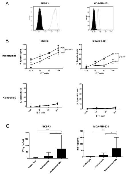
Figure 3.
PSK enhances trastuzumab-mediated ADCC against SKBR3 and MDA-MB-231 breast cancer cells. (A) Expression of HER2 on SKBR3 and MDA-MB-231 cells. The cells were stained with PE conjugated anti-human HER2 (empty histogram) or isotype control (filled histogram). (B) Percentages of specific lysis of trastuzumab- or control IgG-coated SKBR3 and MDA-MB-231 target cells. Shown are mean±SD of triplicate wells at different E:T ratios. ■ indicates PSK-stimulated effector PBMC; ● indicates control PBS-treated effector cells. PBMC were treated with PSK (10 μg/ml) in RPMI for 72 hr before the initiation of cytolytic assay. Difference between PBS and PSK group at different E:T ratio were analyzed using ANOVA. Similar results were obtained using PBMC from 5 different donors as summarized in Supplemental Table. (C) IFN-γ levels (mean±SD) in culture supernatant from the 4 hr ADCC incubation. Control IgG: tumor targets were coated with control irrelevant IgG and incubated with PBMC with no prior PSK stimulation; trastuzumab: tumor cells were coated with trastuzumab and then incubated with PBMC with no prior PSK; trastuzumab+PSK: tumor cells were coated with trastuzumab and incubated with PSK (10 μg/ml, 72 hr)-stimulated PBMC. *, p<0.05; ***, p<0.001 by Mann-Whitney test. Results are representative of three independent experiments.

