Figure 5.
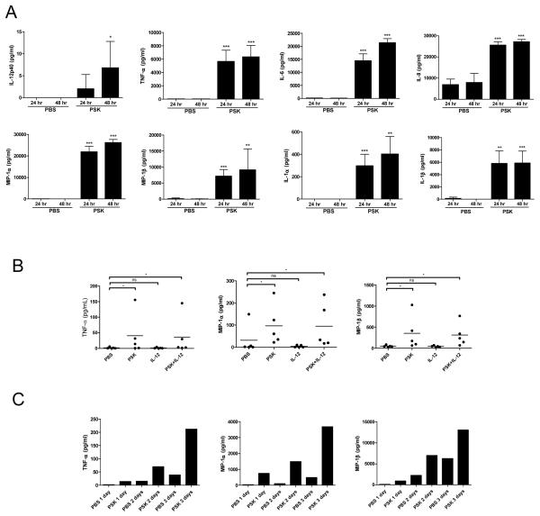
PSK induces the secretion of proinflammatory cytokines and chemokines by PBMC and NK cells. (A) The levels of IL-12p40, TNF-α, IL-6, IL-8, MIP-1α, MIP-1β, IL-1α, and IL-1β in culture supernatant from PBMC treated with PSK (100 μg/ml) or control PBS for 24 or 48 hr, as determined in Luminex analysis. *, p<0.05; **, p<0.01; ***, p<0.001 between PSK and PBS group at the same time point using two-tailed Student t test. Shown are mean±SD of results from 3 independent donors. (B) Shown are levels of TNF-α, MIP-1α, and MIP-1β in culture supernatant from purified NK cells treated with PSK (100 μg/ml), IL-12 (1 ng/ml), or PSK+IL-12. Each data point represents response from an individual donor (N=5). (C) Time course of TNF-α, MIP-1α, and MIP-1β induction by PSK in one donor.

