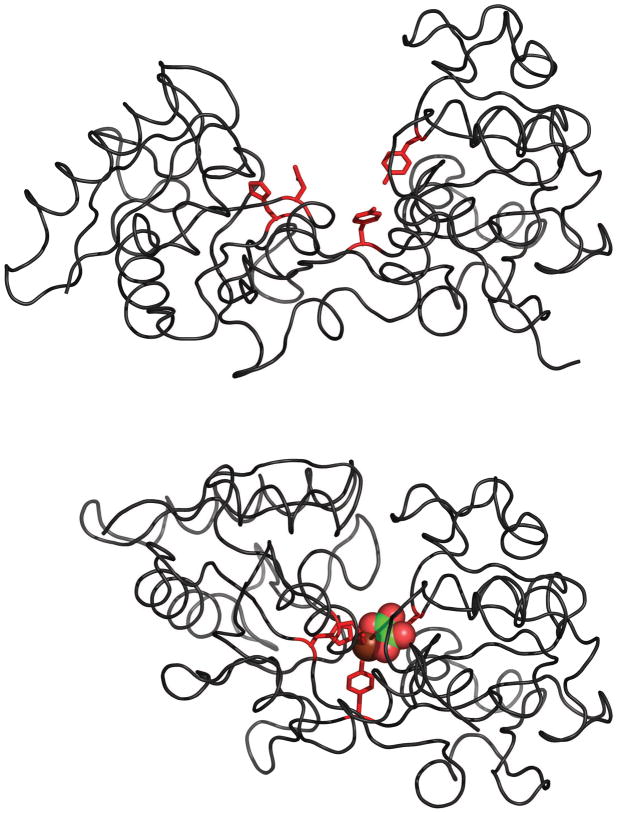
Figure 1.
Crystal structures of the apo- (top) and holo-forms (bottom) of human serum transferrin showing details of iron binding: side chains coordinating Fe3+ are colored in red; both ferric ion and the synergistic anion are presented with space-fill models, and are colored by elements (Fe, chocolate red; C, green; and O, red). The PDB accession numbers are 1BTJ (apo-form [65]) and 1A8E (holo-form [66]).

