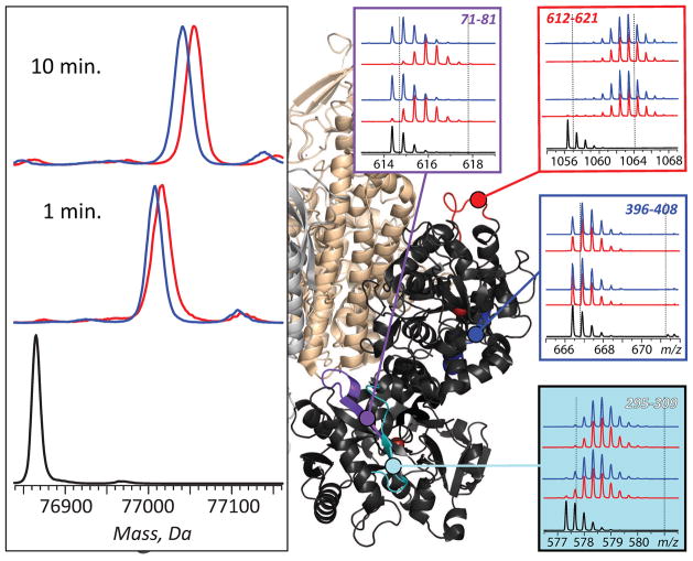
Figure 12.
Effect exerted by the receptor binding on Tf protection and localization of receptor interface with HDX MS. Left panel: HDX MS of Fe2Tf (global exchange) in the presence (blue) and the absence (red) of TfR. The exchange was carried out by diluting the protein stock solution 1:10 in exchange solution (100 mM NH4HCO3 in D2O, pH adjusted to 7.4) and incubating for a certain period of time as indicated on each diagram followed by rapid quenching (lowering pH to 2.5 and temperature to near 0°C). The black trace shows unlabeled protein. Right panel: isotopic distributions of representative peptic fragments derived from Fe2Tf subjected to HDX in the presence (blue) and the absence (red) of the receptor and followed by rapid quenching, proteolysis and LC/MS analysis. Dotted lines indicate deuterium content of unlabeled and fully exchanged peptides. Colored segments within the Fe2Tf/receptor complex show localization of these peptic fragments (based on the low-resolution structure of Tf/TfR [69]). Adapted with permission from [70].

