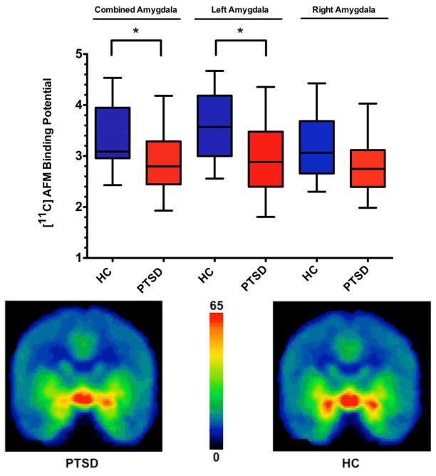
Figure 1. Reduced amygdala [11C]AFM BPND in PTSD compared to healthy subjects.
Upper panel: plot showing [11C]AFM binding potential (BPND) differences in the combined bilateral amygdala region of interest (ROI) and in both left and right amygdala ROI between patients with posttraumatic stress disorder (PTSD) and healthy control subjects (HC). * indicates p<0.05, two-tailed.
Lower panel: Averaged [11C]AFM positron emission tomography (PET) images (coronal view) illustrate reduced amygdala distribution volume in PTSD (left) relative to HC (right).

