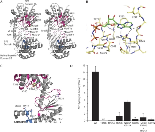
Figure 3.
Conserved motifs and mutational analysis. (A) Stereo view of the structure of RIG-ISF2 with conserved functional motifs shown. (B) Close-up view of the adenosine 5′-(β,γ-imido)triphosphate (AMP-PNP; colour-coded sticks: green, carbon; red, oxygen; blue, nitrogen; orange, phosphorus)-binding moiety with highlighted hydrogen bonds of the adenine recognition site. The protein is shown as colour-coded sticks with yellow carbons. (C) Close-up view of the motifs mutated in this study. Mutated side chains are annotated and shown as sticks. (D) ATP hydrolysis activity of RIG-I SF2 domain mutants. Mixed E374Q+R731A represents a 1:1 mixture of single mutants E374Q and R731A. Plotted bars: mean±s.d. (n=3–6). ND, not detectable; RIG-I, retinoic acid inducible gene I; SF2, superfamily 2; WT, wild type.

