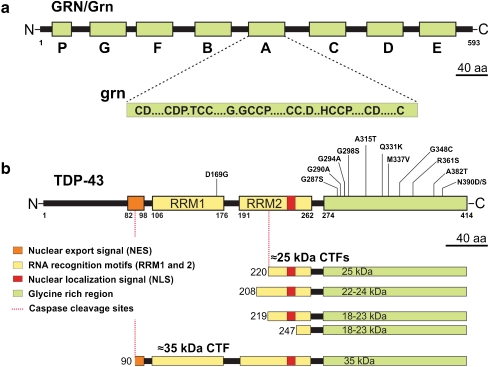
Fig. 1.
Progranulin and TDP-43 structure and processing. a The top part of the figure represents the progranulin protein (human GRN; rodent Grn) and the bottom part shows the consensus sequence of the processed granulin peptides (grn); A–G represent granulin peptides and p represents paragranulin. b Structure of TDP-43 showing different domains of the protein and select mutations mostly clustered in the glycine-rich C terminus of TDP-43. The lower panel shows TDP-43 processing into ∼35-kDa and different ∼25-kDa species identified or speculated to exist in human FTLD/ALS patients. Drawn to scale

