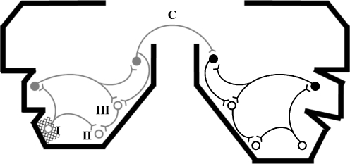
Fig. 2.
A pathophysiological model of semantic dementia. The schematic represents a distributed neural network in both temporal lobes, linked via commissural fibres (C); the temporal lobes are outlined in coronal section (compare Fig. 1) and stylised ‘neurons’ represent different elements of the network. In this representation, the ‘left’ temporal lobe is the more affected (atrophic) but the model does not assume a particular lateralisation. An initial (stochastic) neurodegenerative event (here indicated as cross-hatching at location I) propagates trans-synaptically through the network. The schematic shows a single instigating neurodegenerative focus; however, this could be multifocal. The neurodegenerative process is assumed to be ‘diluted’ as it traverses successive synaptic relays (here coded as grey within the initially maximally affected temporal lobe, and black in the contralateral temporal lobe): though the instigating molecular lesion has not been characterised precisely, such a dilution could in principle result from spread of a toxic molecule or loss of function effects (e.g. synaptic dysregulation or loss of trophic support). Trans-synaptic processes are assumed to act bidirectionally (i.e. potentially both anterograde and retrograde); however, in general there will be a net ‘direction’ of effects according to whether the triggering event is local or remote from the involved network element. The overall result of the process is a gradient of neural damage across the network. At a given disease stage, areas with high synaptic convergence (locations II and III) are more severely affected by the neurodegenerative process (represented as unfilled circles), establishing a gradient of atrophy within the temporal lobe. In addition, there is an inter-hemispheric gradient of damage manifesting as asymmetric temporal lobe atrophy. A precise mapping of commissural pathways between homologous areas in opposite temporal lobes and mirrored network connectivity patterns in each temporal lobe are together assumed to recapitulate the sequence of regional atrophy evolution in the initially less affected temporal lobe

