
Fig. 2.
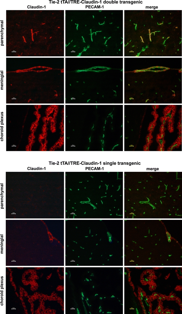
Expression of claudin-1 in the brains of Tie-2 tTA//TRE-claudin-1 double and single transgenic mice. Immunofluorescence staining for claudin-1+ (red) and PECAM-1+ (green) in frozen brain sections of double transgenic Tie-2 tTA//TRE–claudin-1 mice and single transgenic littermates from line 23949 is shown. Expression of claudin-1 was detected in PECAM-1 + parenchymal blood vessels in the brain of Tie-2 tTA//TRE-claudin-1 double transgenic mice but not in single transgenic littermates. In addition, diffuse claudin-1 immunostaining was detected in PECAM-1+ vascular endothelial cells within the leptomeninges and in cell-junctions of choroid plexus epithelial cells in Tie-2 tTA//TRE-claudin-1 double transgenic and single transgenic mice. Scale bar 20 μm