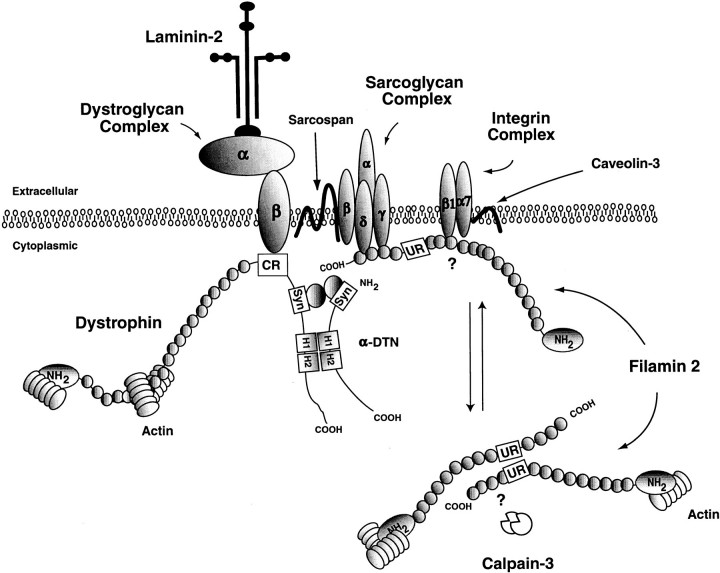
Figure 10.
LGMD model. Shown is a schematic representing some proteins involved in muscular dystrophy and how they might interact with FLN2. Depending on the phosphorylation state of FLN2, it may be present at the membrane or interacting with the actin cytoskeleton. At the membrane, FLN2 interacts with γ- and δ-sarcoglycan and possibly with β1 integrin. One set of receptor signals may recruit FLN2 to the membrane, whereas another set of signals allows FLN2 to translocate back to the actin cytoskeleton. Also depicted in this diagram is calpain-3, which may help to regulate FLN2 levels within the cell.

