
Figure 5.
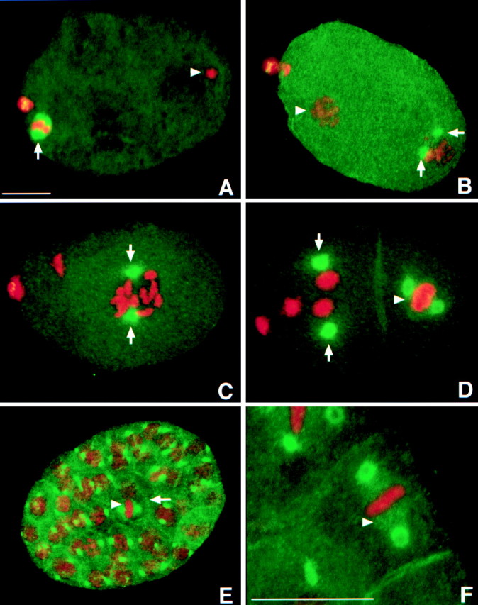
LIN-5 localizes in a cell cycle-dependent manner to the meiotic spindle, mitotic centrosomes, and kinetochore microtubules. In addition, LIN-5 is present in the cytoplasm and at the cell periphery. Staining with LIN-5–specific antibodies is indicated in green, DNA staining with PI in red. A, Fertilized embryo in meiosis II. LIN-5 was localized to the meiotic spindle (arrow). The condensed sperm pronucleus is visible at the posterior of the embryo (arrowhead). Some LIN-5 protein was expelled with the polar bodies that are visible in A–D as large red dots at the anterior (left in all panels) of the embryo. B, One-cell embryo at the time of migration of the maternal pronucleus. LIN-5 was located at the duplicated centrosomes of the decondensed sperm pronucleus (arrows) and was no longer detected at the oocyte pronucleus (arrowhead). C, LIN-5 remained associated with the centrosomes (arrows) during nuclear migration, rotation, and formation of the first mitotic spindle. D, LIN-5 is detected at the centrosomes (arrows) and kinetochore microtubules (arrowhead) of this two-cell embryo. E, LIN-5 is detected at the centrosomes, metaphase spindle (arrowhead), and cell cortex (arrow) of mitotic cells throughout embryogenesis. F, Magnification of a multicellular embryo to illustrate the localization of LIN-5 at the kinetochore microtubules (arrowhead) in metaphase cells. Bars, ∼10 μm.