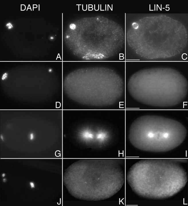
Figure 6.
LIN-5 localization is disrupted by treatment with nocodazole. Immunofluorescence micrographs of untreated (A–C and G–I) and nocodazole-treated (D–F and J–L) wild-type embryos stained with DAPI (left), antitubulin antibodies (middle), or anti–LIN-5 antibodies (right) during meiosis (A–F) and mitosis (G–L). In embryos with depolymerized microtubules, LIN-5 did not localize to the meiotic spindle, centrosomes, and metaphase spindle. Anterior is to the left. Bars, ∼10 μm.

